The Birth of Ultrasound
Chapter Three
What did the world's first ultrasound look like?
Introduction
In 1958, something remarkable happened in Glasgow.
For the first time in history, doctors could look safely inside the womb and see a living baby before birth.
Today, ultrasound scans are a familiar part of pregnancy. Every year, millions of families around the world experience the excitement of seeing their baby for the first time.
But before that moment in Glasgow, it had never been possible.
The images were simple.
The equipment was experimental.
The possibilities were endless.
Film 3: The Birth of Ultrasound
This film tells the story of the world's first successful medical ultrasound scans. Through archive photographs, historic footage and expert insight, discover how Professor Ian Donald and Tom Brown transformed an experimental machine into a medical breakthrough that would change the experience of pregnancy forever.
A Moment That Changed Medicine
After months of experimentation, Professor Ian Donald and Tom Brown were ready to test their new scanner.
The machine looked nothing like the sleek ultrasound systems we know today.
It was large.
Heavy.
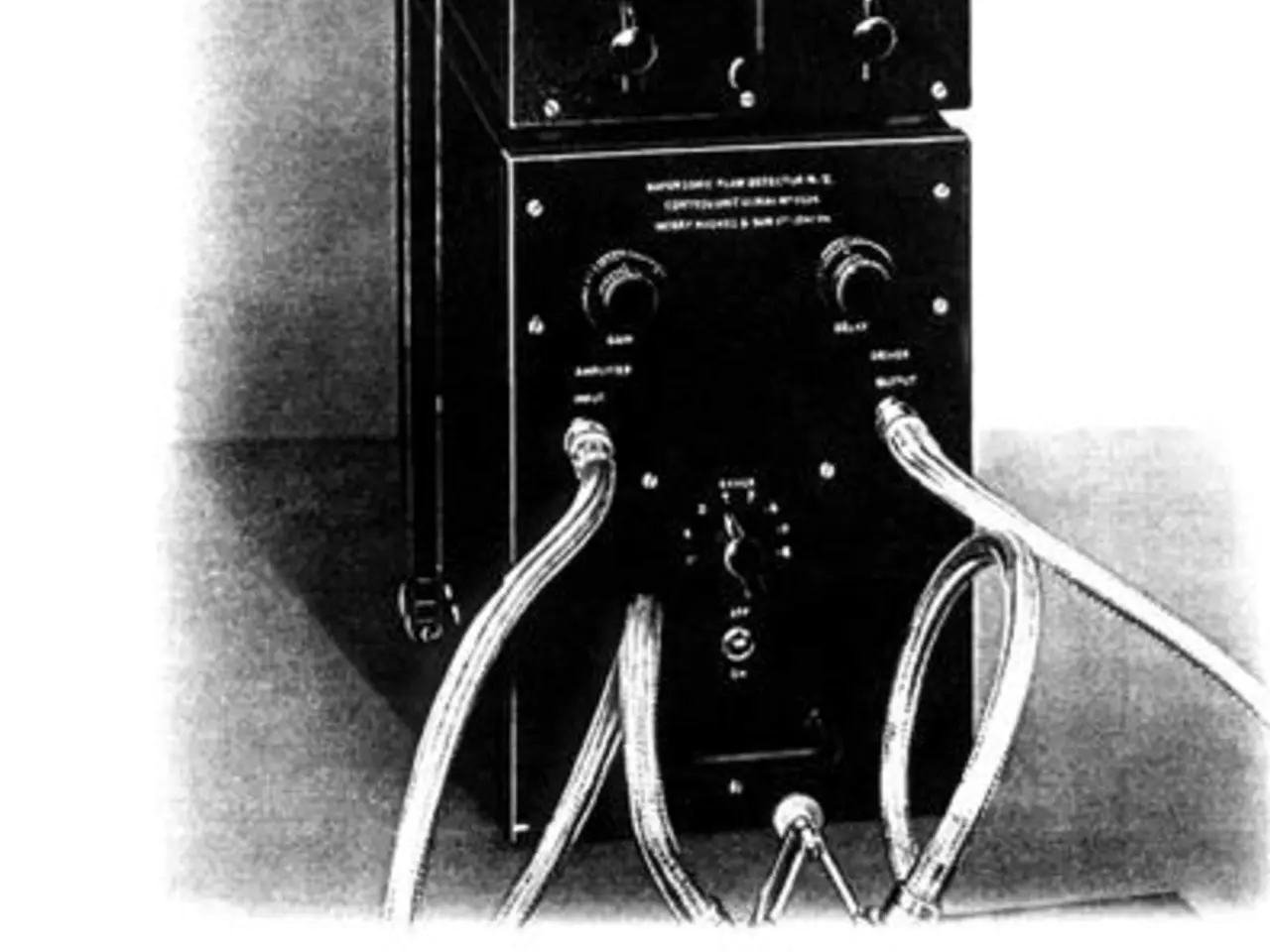
Full of valves, wires and electronics originally designed for industrial engineering.
Yet beneath its metal casing was an entirely new way of understanding the human body.
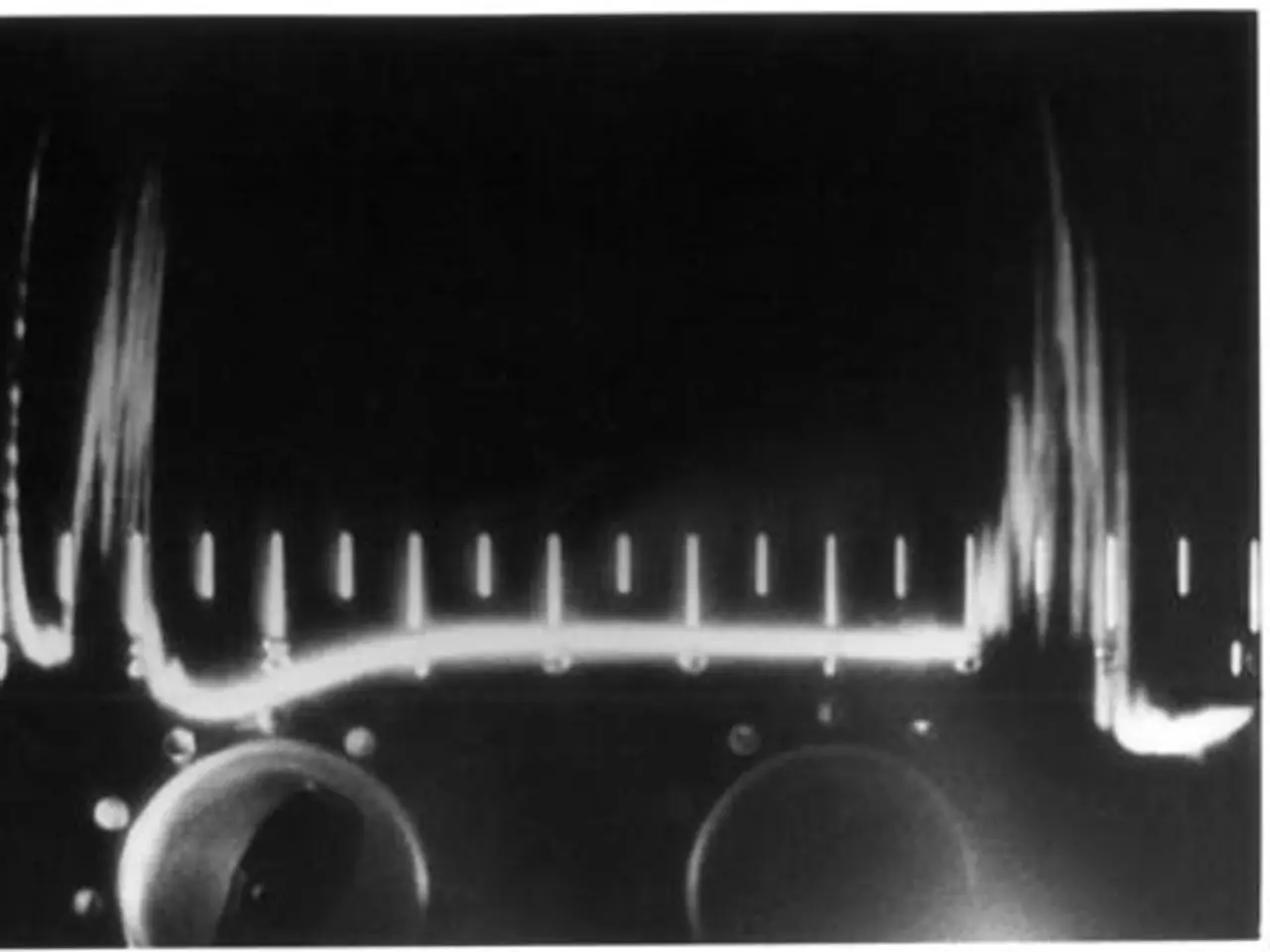
As the ultrasound probe moved across the patient, high-frequency sound waves travelled through the body before bouncing back from different tissues.
Those returning echoes appeared as flickering lines on a green cathode-ray screen.
To modern eyes, the image seems almost impossible to interpret.
To Donald and Brown, it was extraordinary.
For the first time, they could distinguish structures hidden safely inside the body without surgery or harmful radiation.
Medicine would never be the same again.
Seeing Life Before Birth
The greatest impact of this new technology was felt in maternity care.
Until this point, doctors relied almost entirely on physical examination and experience to understand what was happening during pregnancy.
Ultrasound changed that forever.
For the first time, parents and clinicians could see evidence of life before birth.
The images were grainy.
They lacked the detail we expect today.
But they answered questions that had previously been impossible to answer safely.
How was the baby growing?
Was the pregnancy progressing normally?
Could complications be identified earlier?
Within the first year of clinical use, around 250 pregnant women were scanned using the pioneering Glasgow technology.
Each scan represented another step towards a future where ultrasound would become routine healthcare.
From Experiment to Evidence
Early ultrasound did far more than produce remarkable images.
It proved that sound could become one of medicine's safest and most valuable diagnostic tools.
Researchers continued refining the technology, improving image quality and developing new ways to interpret the echoes returning from inside the body.
What began as an experimental project in Glasgow quickly attracted international attention.
Hospitals around the world wanted to understand how this new technology worked.
The journey from shipyard innovation to global medical practice had begun.
Interactive 3:Virtual Ultrasound
Learning Goal
Experience one of the world's first ultrasound examinations and discover how doctors learned to interpret the earliest medical images created using sound.
Before You Begin
Imagine you are standing beside Professor Ian Donald in Glasgow during the late 1950s.
The prototype scanner is ready.
No one knows exactly what will appear on the screen.
As you move the ultrasound probe across the patient's abdomen, sound waves travel through the body before returning as echoes.
Watch carefully as the image slowly appears.
Although it may look very different from modern ultrasound scans, this simple display represented one of the greatest breakthroughs in medical history.
Can you identify the structures revealed by the returning echoes?
What Have You Discovered?
The image you created may seem unclear compared with today's technology.
That is exactly what makes it so remarkable.
For the first time, doctors could safely look inside the body without making an incision or exposing patients to harmful radiation.
Every modern ultrasound scan, from pregnancy and heart imaging to emergency medicine, can trace its origins back to discoveries made using machines like this.
What began as flickering green lines became one of the most important diagnostic tools in healthcare.
Explore the Original Innovation
Parts of the world's first ultrasound scanner are carefully preserved by The Hunterian in Glasgow.
Although the complete machine could not be retained because some components contained asbestos, these surviving pieces tell the story of one of Scotland's greatest scientific achievements.
Historic photographs, original equipment and archive material continue to help researchers, students and visitors understand how ultrasound was born.
Did You Know?
The first ultrasound images appeared as simple green lines on a cathode-ray display.
Today's scanners create highly detailed 2D, 3D and even 4D images in real time, but they still rely on exactly the same scientific principle: interpreting echoes returned by high-frequency sound waves.
Reflect
Imagine seeing one of these images for the very first time in 1958.
You have never seen a baby before birth.
You don't know what to expect.
How do you think that moment might have changed the relationship between parents, clinicians and the unborn child?
Continue Your Journey
Ultrasound didn't just change medicine.
It changed people's lives.
In the next chapter, you'll hear from the women, families, midwives and clinicians who experienced this remarkable technology during its earliest years and discover how a scientific breakthrough became a deeply human story.
Continue to Chapter Four